


MTW European Type Trapezium Mill
Input size:30-50mm
Capacity: 3-50t/h
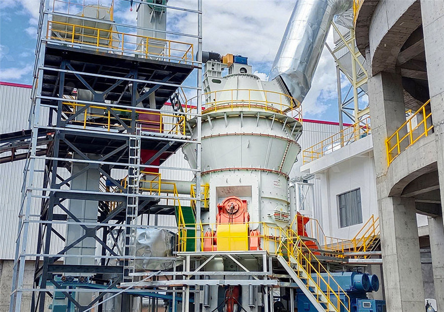
LM Vertical Roller Mill
Input size:38-65mm
Capacity: 13-70t/h
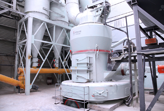
Raymond Mill
Input size:20-30mm
Capacity: 0.8-9.5t/h
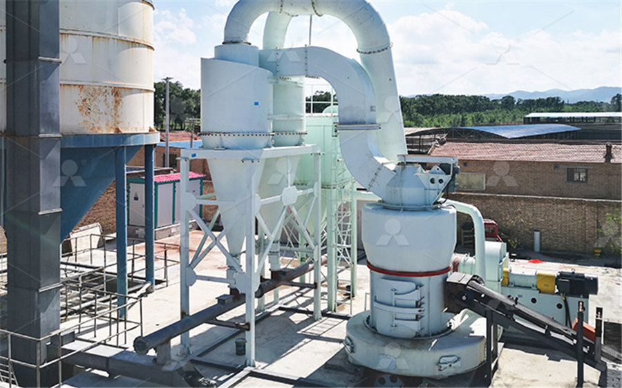
Sand powder vertical mill
Input size:30-55mm
Capacity: 30-900t/h
LUM series superfine vertical roller grinding mill
Input size:10-20mm
Capacity: 5-18t/h
MW Micro Powder Mill
Input size:≤20mm
Capacity: 0.5-12t/h

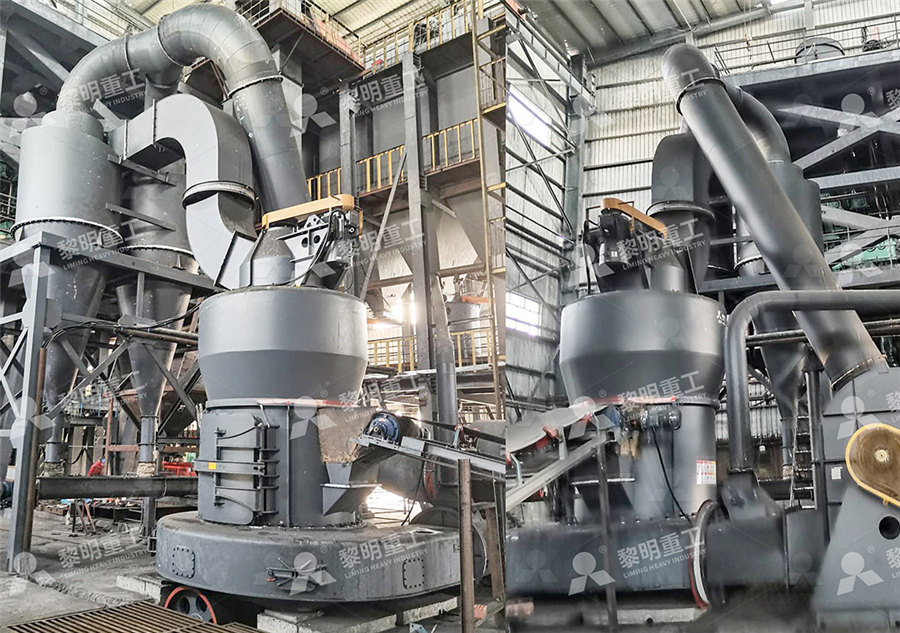
LM Vertical Slag Mill
Input size:38-65mm
Capacity: 7-100t/h

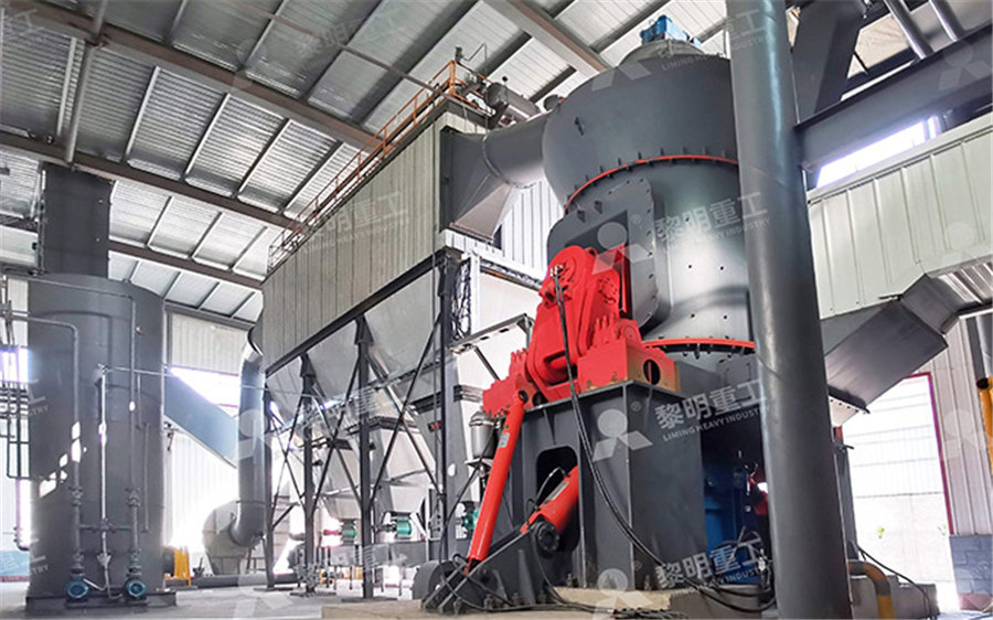
LM Vertical Coal Mill
Input size:≤50mm
Capacity: 5-100t/h
TGM Trapezium Mill
Input size:25-40mm
Capacity: 3-36t/h

MB5X Pendulum Roller Grinding Mill
Input size:25-55mm
Capacity: 4-100t/h
Straight-Through Centrifugal Mill
Input size:30-40mm
Capacity: 15-45t/h
Palatal lobes pictures
.jpg)
4 DENTAL MORPHOLOGY Pocket Dentistry
2024年2月17日 They all have some common characteristics that can help us identify them: a relatively straight incisal edge with a curvature in distal, a relatively rectangular crown, three labial lobes (form the vestibular surface) 2024年10月2日 We have created 114 medical original illustrations of the mouth, the buccal cavity, the bones of the palate, the tongue, the salivary glands and the oral part of the pharynx Anatomy of the oral cavity and the mouth: annotated IMAIOS2023年2月23日 The labial face has three lobes named mamelons, divided by two cervicalincisal groves Due to function, the mamelons tend to become less evident The cervical outline of the Dental Anatomy and Morphology of Permanent Teeth IntechOpenThe palatal root is more broad mesiodistally than buccolingually and ovoidal in shape but normally contains only a single canal Although the palatal root generally appears straight on Maxillary first molar Wikipedia
16 Permanent Anterior Teeth Pocket Dentistry
2015年1月5日 Permanent anterior teeth include the incisors and canines (Figure 161, see Figure 24, 151) All anterior teeth are composed of four developmental lobes: three labial Anatomy of the oral cavity; drawing shows the lip, hard palate, soft palate, retromolar trigone, front twothirds of the tongue, gingiva, buccal mucosa, and floor of mouth Also shown are the Oral Cavity Anatomy: Image Details NCI Visuals Online Cancer2023年11月3日 The oropharyngeal isthmus is surrounded by the soft palate and palatoglossal arches A number of bones contribute to the framework of the oral cavity; these are the paired Oral cavity: Anatomy, tongue muscles, nerves and vessels KenhubBoth grooves improve the aesthetical look of the tooth and, at the same time, separate three lobes These lobes are called the mesial , central and distal lobes The palatal surface Maxillary central incisor Incisors Dental Anatomy

11: The Permanent Maxillary Molars Pocket Dentistry
2015年1月9日 Maxillary First Molar Figures 111 through 1118 illustrate the maxillary first molar from all aspects The crown of this tooth is wider buccolingually than mesiodistally Usually the extra dimension buccolingually 2015年1月9日 Maxillary Central Incisor Figures 61 through 612 illustrate the maxillary central incisor in various aspects The maxillary central incisor is the widest mesiodistally of any of the anterior teeth ()The labial face is less 6: The Permanent Maxillary Incisors Pocket Dentistry2023年7月8日 Palatal Tori (Torus Palatinus) – Treatment, Pictures, Cancer, Cause, Removal, Symptoms July 8, 2023 / Palatal tori, or torus palatinus, refers to benign bony growths that develop along the hard palate They are a common anatomical variation and often discovered during a routine clinical examinationPalatal Tori (Torus Palatinus) – Treatment, Pictures, Cancer, Cause 2015年1月5日 The occlusal surface of permanent posteriors creates an inner occlusal table bordered by the marginal ridges (Figure 173)There are also triangular ridges, which are cusp ridges that descend from the cusp tips toward the central part of the occlusal table (Figure 174)They are so named because the slopes of each side of the ridge are inclined in a way that 17 Permanent Posterior Teeth Pocket Dentistry
.jpg)
Maxillary first molar Wikipedia
The maxillary first molar is the human tooth located laterally (away from the midline of the face) from both the maxillary second premolars of the mouth but mesial (toward the midline of the face) from both maxillary second molars The function of this molar is similar to that of all molars in regard to grinding being the principal action during mastication, commonly known as chewing2024年9月4日 Palatal groove represents a relatively uncommon developmental root anomaly, usually found on the palatal aspect of maxillary incisors While its origin is controversial, its presence predisposes to severe periodontal defects This study aimed to provide a systematic review of the literature focusing on the varied diagnostic techniques and treatment modalities Palatal groove associated with periodontal lesions: a systematic 2021年9月20日 In addition to the distal and the palatal roots, part of the mesial root is also visible from the distal aspect because the distobuccal root is narrow Occlusal Aspect The permanent maxillary first molar is wider on the mesial side as compared to the distal side The tooth is wider mesiodistally on the lingual aspect as compared to the buccal Permanent Maxillary First Molar: Tooth Morphology Made Easy!2023年5月24日 Palatal Tori (Torus palatinus) is a similar condition of a dental tori, but it refers to a bone growth on – on Tongue, Treatment, Causes, Pictures Images, Contagious July 15, 2023 Canker Sores vs Cold Sores – Treatment, Pictures, Contagious, in Throat, on Tonsils, on Roof of Mouth July 11, 2023 Sialadenitis – Treatment Mandibular Tori: Removal, Symptoms, Causes, Surgery Treatment and Pictures

Bifid uvula: Causes, complications, and pictures Medical News
2018年2月11日 A look at bifid uvula, a condition affecting the palate of the mouth Included is detail on complications of the condition and outlook for a bifid uvula2015年1月4日 On the bifurcated root form, one buccal (facial) and one palatal (lingual) root are present The buccal root is larger and longer than the palatal root (see Fig 141, D and E) On the singlerooted form, grooves are usually present lengthwise in the middle of the root, giving the appearance of a root trying to divide itself14: Premolars Pocket Dentistry2021年6月13日 The Palatal outline of the crown is convex in the region of the cingulum and concave at the marginal ridge The curvature of the cervical line (cementoenamel junction) is towards the incisal aspect The Maxillary central incisor has the greatest curvature of the cervical line on the mesial aspect than any other toothPermanent Maxillary Central Incisor Dental Education Hub2018年8月1日 They developed from four lobes, three labially and one lingually (palatally), the lingual (palatal) lobe being represented by the cingulum Each labial lobe of the incisor Permanent Maxillary and Mandibular Incisors
First maxillary molar (part 1) Molars Dental Anatomy
The first maxillary molar is the most significant tooth of all maxillary molar teeth It has a rhomboid shape, and its mesiodistal length is slightly smaller than the buccolingual length The buccal mesiodistal length of the first maxillary molar is narrower than the same palatal length The cervical distobuccal line angle is concave and rounded, and it is a common site for plaque The uvula (pl: uvulas or uvulae), also known as the palatine uvula or staphyle, is a conic projection from the back edge of the middle of the soft palate, composed of connective tissue containing a number of racemose glands, and some muscular fibers [1] [2] It also contains many serous glands, which produce thin saliva [3]It is only found in humansUvula Wikipedia2022年7月8日 What are some conditions that affect tonsils? There are a few different conditions that can affect your tonsils The most common is tonsillitis — an infection of the tonsils Bacteria and viruses can cause tonsillitis, and the infection can Tonsils: Anatomy, Definition Function Cleveland Clinic2023年10月30日 Structure All eight incisors share a general tooth anatomy, consisting of a crown that is continued by the rootThe crown is covered by enamel, while the root is covered by cementumThe body of the tooth consists almost entirely of dentine which surrounds a central pulp cavityThis cavity expands into a pulp chamber, which narrows at the end into the root canalIncisors: Definition, shape, function Kenhub
Cingulum eAnatomy IMAIOS
The cingulum refers to the portion of the teeth (anterior teeth (incisors and canines)), occurring on the lingual or palatal aspects, that forms a convex protuberance at the cervical third of the anatomic crown It represents the lingual or palatal developmental lobe of these teethSearch from Parietal Lobe stock photos, pictures and royaltyfree images from iStock For the first time, get 1 free month of iStock exclusive photos, illustrations, and more2,800+ Parietal Lobe Stock Photos, Pictures RoyaltyFree The central lobe is better developed than the mesial and distal These lobes are separated by the mesial and distal developmental grooves Also, the maxillary canine has a very noticeable notch at the cementoenamel junction The palatal surface shows such structures as the cingulum, mesial and distal marginal ridges and lingual ridgeMaxillary canine Canines Dental Anatomy2013年2月12日 It is also stated that maxillary premolars and mandibular first premolar develop from four lobes (mesial, distal, buccal, and palatal), whereas mandibular second premolar which often has two lingual cusps develop from five lobes (mesial, buccal, distal, mesiolingual, and distolingual lobes) Maxillary and Mandibular First Premolars Showing Three‐Cusp
.jpg)
Dental Anatomy and Occlusion: Mandibular Incisors—Flipped
This dental anatomy module is the second in a series that develops skills in analyzing the morphology, function, anomalies, and development of human teeth Learning the visual details associated with teeth has often proven difficult using the Find Palatal Expander stock images in HD and millions of other royaltyfree stock photos, illustrations and vectors in the Shutterstock collection Thousands of new, highquality pictures added every dayPalatal Expander Photos, Images Pictures ShutterstockSearch from Palate stock photos, pictures and royaltyfree images from iStock For the first time, get 1 free month of iStock exclusive photos, illustrations, and more Video Back Removing taking off dental braces and teeth palatal 8,000+ Palate Stock Photos, Pictures RoyaltyFree 2015年1月9日 Buccal Aspect From the buccal aspect, the crown is roughly trapezoidal (see Figure 416, C)The crown exhibits little curvature at the cervical line The crest of curvature of the cervical line buccally is near the center of the root buccally (see Figure 92 and Figures 97 through 99) The mesial outline of the crown is slightly concave from the cervical line to the 9: The Permanent Maxillary Premolars Pocket Dentistry

Exploring the Phonological Process of Palatalization Kidoneo
In this case, the nonpalatal consonant “k” was transformed into the palatal consonant “ch” Palatalization is not limited to the transformation of nonpalatal consonants into palatal consonants It can also involve the transformation of alveolar fricatives into palatal fricativesSearch from Lobe stock photos, pictures and royaltyfree images from iStock For the first time, get 1 free month of iStock exclusive photos, illustrations, and more Video Back Videos home; Cerebral cortex lobes in color profile view with body isolated on white background accurate 3D rendering illustration Human brain anatomy, neurology14,800+ Lobe Stock Photos, Pictures RoyaltyFree ImagesThe aggregate of cells which eventually form a tooth are derived from the ectoderm of the first branchial arch and the ectomesenchyme of the neural crest [7] As in all cases of tooth development, the first hard tissue to begin forming is dentin, with enamel appearing immediately afterwards [8] [page needed]The deciduous maxillary central incisor begins to undergo Maxillary central incisor Wikipedia2024年2月27日 A torus palatinus is a growth that forms on the roof of the mouth They may be present at birth or develop later They tend to be harmless but can cause complicationsTorus palatinus: Pictures, symptoms, and treatments Medical
.jpg)
Speech anatomy for English pronunciation • icSpeech
Sounds that are made with the hard palate are called palatal sounds, for example /j/ Soft Palate (velum) The soft palate or velum is the soft portion of the roof of the mouth, lying behind the hard palate The velum performs two important roles in speech:Find Brain Lobes stock images in HD and millions of other royaltyfree stock photos, illustrations and vectors in the Shutterstock collection Thousands of new, highquality pictures added every day12,527 Brain Lobes RoyaltyFree Images, Stock Photos Pictures 2024年2月27日 Palatal expanders have high success rates in children and adults Studies have shown a success rate of between 84 and 88% 8,9 One study noted complications are much less likely if palatal expansion occurs 4 Types of Palate Expanders (Uses, Side EffectsDefinition of palatal adjective in Oxford Advanced Learner's Dictionary Meaning, pronunciation, picture, example sentences, grammar, usage notes, synonyms and morepalatal adjective Definition, pictures, pronunciation and usage
Lobes of the brain: Structure and function Kenhub
2023年10月30日 Key facts about the lobes of the brain; Frontal lobe Location: Corresponds to the frontal bone; Anterior to the parietal lobe (separated by central sulcus) and superior and anterior to the temporal lobe (separated by Oral tori are common exostoses with a prevalence of about 20–30% in the general USA population [] Torus palatinus is more common in women and in people of Asian and Inuit ancestry [2,3] The pathogenesis is not wellunderstood, and appears to be a complex interplay of occlusive (biting) forces, genetics, and environmental factors[3–5] Medical conditions Images in medicine: torus palatinus PMC PubMed Central Definition of palatal noun in Oxford Advanced American Dictionary Meaning, pronunciation, picture, example sentences, grammar, usage notes, synonyms and morepalatal noun Definition, pictures, pronunciation and usage notes Fun educational facts about the brain and the four lobes HOME NEW POPULAR RESOURCES Brain Activities Other our occipital lobe also has to combine information from both eyes so that we only see one picture That's A LOT of work for one lobe! References: Kolb, B, Whishaw, I Q, Campbell Teskey, G (2000) An introduction to brain and Brain Facts: The Four Lobes
.jpg)
Morphology of Permanent Molars LWW
The number of lobes forming molars is one per cusp, including the cusp of Carabelli See Table 51 for a summary of the number of lobes forming first and second molars 3 CROWN TILT THAT DISTINGUISHES MAXILLARY FROM MANDIBULAR MOLARS When mandibular molar crowns are examined from the proximal views, the crowns appear to be tilted lin2021年8月16日 Palatal Aspect The crown and the root both are narrow mesiodistally as compared to the labial aspect Therefore, part of the mesial and distal surfaces of both crown and root are visible from the palatal aspect The mesial and distal marginal ridges of the maxillary canine are very well developedPermanent Maxillary Canine Dental Education Hub2023年3月19日 Most people with palatal tori are over the age of 30 Genetics Research indicates that you’re more likely to develop palatal tori if your biological parents, grandparents or siblings have the condition What are the complications of torus palatinus (palatal tori)? Palatal tori aren’t harmful or dangerousTorus Palatinus (Palatal Tori) Cleveland ClinicDefinition of palatal noun in Oxford Advanced American Dictionary Meaning, pronunciation, picture, example sentences, grammar, usage notes, synonyms and morepalatal noun Definition, pictures, pronunciation and usage notes